Program
Breakfast
Introduction
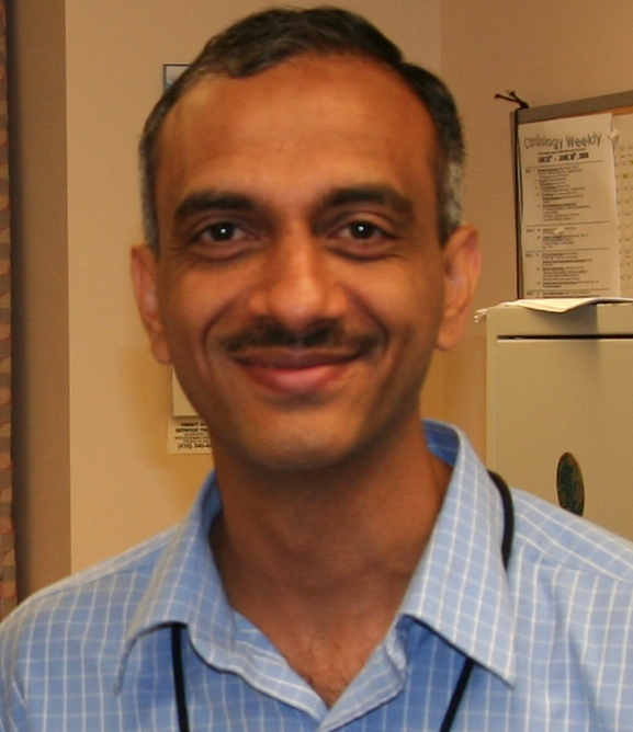
Raja Selvaraj
A quick introduction, setting out the key elements of the program. Objectives, important design aspects, feedback, evaluation.
Essential anatomy for EP
Raja Selvaraj
External anatomy
Internal anatomy of RA, RV, LA and LV
Fluoroscopic anatomy for EP
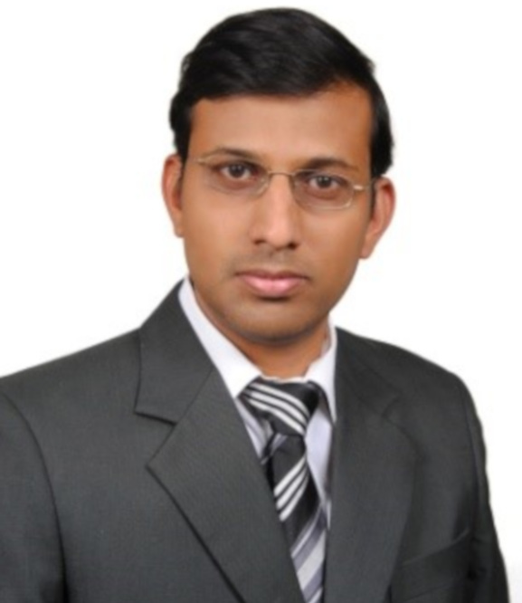
John Roshan
Basic views and anatomy in each view. Applied fluoroscopic anatomy for each chamber. Useful views while mapping different arrhythmias.
ECG in relation to EP - Sinus
John Roshan
ECG in sinus rhythm - Preexcitation, Ebstein anomaly, DAVNP
Coffee break
ECG in relation to EP - Tachycardia
Raja Selvaraj
ECG findings of relevance to EP study in patients with narrow and wide complex tachycardia.
EP lab hardware
John Roshan
Diagnostic and ablation catheters. HD mapping catheters
Sheaths
EP Signals and measurements
Raja Selvaraj
EP lab set up
Filtering and amplification
Bipolar and unipolar signals
Amplifier set up, page set up
Views - real time, review, triggered
Lunch
Stimulator and Pacing protocols
John Roshan
Stimulator, setting up sensing different pacing protocols
Biophysics of RF - Basics
Raja Selvaraj
Biophysics principles
Impact of electrode size, power, time and contact force
Irrigation, different irrigants
Advanced RF biophysics, biophysics of Cryo and PFA
John Roshan
Biophysics of bipolar ablation / simultaneous unipolar ablation
Biophysics of cryo ablation
Biophysics of PFA
Coffee break
Normal electrophysiologic responses
Raja Selvaraj
Normal response with ventricular pacing
Normal response with atrial pacing
Narrow QRS tachycardia - Evaluation in sinus rhythm
John Roshan
Distinguishing nodal vs extranodal VA conduction. Identifying dual AV nodal physiology. Preexcitation patterns and response to extrastimuli. Parahisian pacing
Narrow QRS tachycardia - Evaluation during tachycardia part I
Raja Selvaraj
Tachycardia measurements, VA relationship, VA interval and atrial activation sequence
VOP to distingish mechanisms
End of day - Beach and dinner
Breakfast
Narrow QRS tachycardia - Evaluation during tachycardia part II
John Roshan
His refractory PVC, AH during tachycardia, VA linking, simultaneous AV pacing
AVNRT - Mechanism and Diagnosis
Raja Selvaraj
AVNRT types and mechanism
Basic diagnosis - tachycardia, pacing maneuvers
DD and how to differentiate - AVRT, AT, NV/NF AVRT
AVNRT - Mapping and ablation
Raja Selvaraj
Identifying the operative slow pathway - resetting response
Mapping approach - fluoro, EGM, 3D
Ablation - power, monitoring VA conduction
AV accessory pathways - Evaluation and diagnosis
John Roshan
Tachycardia characteristics
Diagnosing AVRT, DD
Coffee break
AV accessory pathways - Mapping and ablation
John Roshan
General principles of mapping accessory pathways.
Mapping and ablation of septal pathways
Mapping and ablation of right-sided pathways.
Mapping and ablation of left-sided pathways
Atypical accessory pathways - Evaluation and diagnosis
Raja Selvaraj
CS accessory pathways
Atrial appendage to ventricle pathways
atriofascicular - only brief mention
nodoventricular / nodofascicular
Fasciculoventricular
Atriofascicular accessory pathways - Mapping and ablation
John Roshan
Atriofascicular AP - Anatomy and EP characteristics
Antidromic AVRT - evaluation
Mapping and ablation
Lunch
Atrial tachycardia and flutter - Evaluation and diagnosis
Raja Selvaraj
Terminology
AT - identifying chamber
A flutter - entrainment, mapping
Atrial flutter - Mapping and ablation
John Roshan
Mapping and ablation of typical flutter
Establishing bidirectional block
Mapping and ablation of atypical flutters
Mitral isthmus ablation
Case in the box - SVT I
JIPMER faculty
Coffee break
Wide QRS tachycardia - ECG and evaluation in sinus
John Roshan
ECG - Identifying common wide QRS tachycardias. Clues during atrial and ventricular pacing.
Wide QRS tachycardia - Evaluation in tachycardia
Raja Selvaraj
Differentiation of SVT, VT, and preexcited tachycardia. Identification of bundle branch reentry.
Case in box 2 - WCT
CMC faculty
End of day and dinner
Breakfast
Idiopathic RVOT VT
John Roshan
ECG morphology
RV versus LV OT locations
Mapping and ablation
Idiopathic LV fascicular VT
Raja Selvaraj
Clinical presentation and ECG
Variants and proposed circuits
Mapping and ablation
Delegate evaluation
Raja Selvaraj and John Roshan
10 questions each
Coffee break and Group Photo